Introduction to Animal Cells
Animal cell reading comprehension and coloring – Animal cells are the fundamental building blocks of animal tissues and organs. They are eukaryotic cells, meaning they possess a membrane-bound nucleus and other organelles. Understanding their structure and function is crucial to comprehending the complexities of animal life.Animal cells perform a wide range of functions essential for survival, including nutrient uptake and processing, energy production, waste removal, and cell division.
These functions are carried out by specialized structures within the cell, known as organelles. The coordinated actions of these organelles maintain cellular homeostasis and contribute to the overall health of the organism.
Animal Cell Organelles and Their Roles
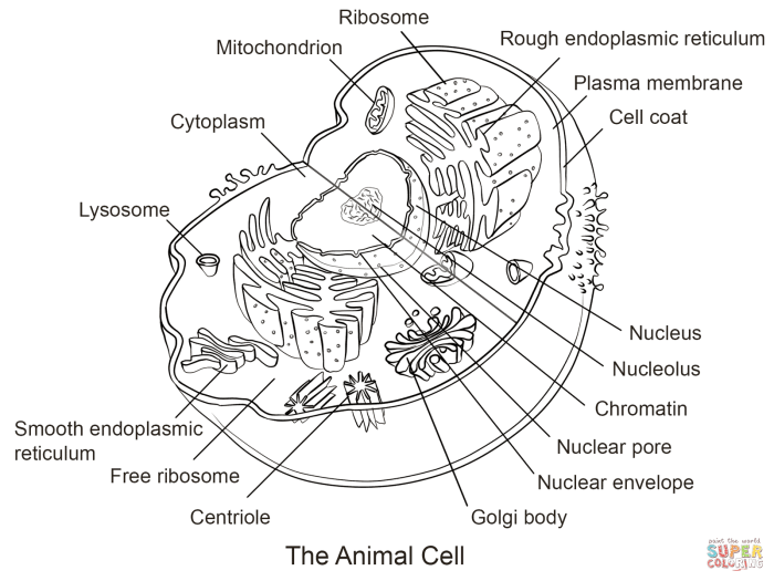
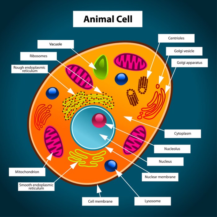
The following table details the major organelles found in animal cells, their functions, descriptions, and illustrative representations.
| Organelle Name | Function | Description | Illustration Description |
|---|---|---|---|
| Nucleus | Contains the cell’s genetic material (DNA) and controls cell activities. | A large, round structure usually located near the center of the cell. It is enclosed by a double membrane called the nuclear envelope. | A large, centrally located circle containing smaller, darker circles representing the nucleolus and chromatin. The circle is surrounded by a double-lined membrane. |
| Mitochondria | Generate energy (ATP) through cellular respiration. | Rod-shaped or oval organelles with a folded inner membrane (cristae). Often referred to as the “powerhouses” of the cell. | Bean-shaped structures with internal folds depicted as wavy lines within the bean shape. |
| Ribosomes | Synthesize proteins. | Small, granular structures found free in the cytoplasm or attached to the endoplasmic reticulum. | Small dots scattered throughout the cytoplasm and also attached to a network of interconnected tubes and sacs (the endoplasmic reticulum). |
| Endoplasmic Reticulum (ER) | Synthesizes lipids, modifies proteins, and transports materials. | A network of interconnected membranes forming sacs (cisternae) and tubules. There are two types: rough ER (with ribosomes attached) and smooth ER (without ribosomes). | A network of interconnected tubes and sacs, some studded with small dots (ribosomes) representing rough ER and some smooth, representing smooth ER. |
| Golgi Apparatus (Golgi Body) | Processes and packages proteins and lipids for secretion or transport within the cell. | A stack of flattened, membrane-bound sacs (cisternae). | A stack of flattened sacs resembling pancakes, often with small vesicles budding off from the edges. |
| Lysosomes | Break down waste materials and cellular debris. | Small, membrane-bound sacs containing digestive enzymes. | Small, round sacs containing smaller, darker dots representing the digestive enzymes. |
| Cell Membrane | Regulates the passage of substances into and out of the cell. | A thin, flexible outer boundary of the cell. It is selectively permeable. | A thin, continuous line outlining the entire cell, suggesting a membrane surrounding the organelles. |
| Cytoplasm | The jelly-like substance filling the cell, containing organelles and cytosol. | The fluid-filled space within the cell membrane, excluding the nucleus. | The clear space within the cell membrane, containing all the other depicted organelles. |
| Centrioles | Play a role in cell division. | Paired cylindrical structures located near the nucleus. | Two small, cylindrical structures located close to the nucleus, often depicted at right angles to each other. |
Animal Cell Structure and Function

Animal cells, the fundamental building blocks of animals, are complex structures containing various organelles, each with a specific role in maintaining cellular life. Understanding their structure and function is crucial to grasping the intricacies of animal biology and physiology. This section delves into the individual components of the animal cell and their collaborative efforts in key cellular processes.
Animal cells are eukaryotic cells, meaning they possess a membrane-bound nucleus housing their genetic material. Beyond the nucleus, a variety of other organelles work together to perform essential functions, including energy production, protein synthesis, and waste removal. The coordinated actions of these organelles ensure the cell’s survival and contribute to the overall health of the organism.
Comparison of Animal Cell Organelles
The following table compares the structures and functions of several key animal cell organelles:
| Organelle | Structure | Function |
|---|---|---|
| Nucleus | Large, membrane-bound organelle containing DNA | Controls gene expression, regulates cellular activities |
| Ribosomes | Small, RNA-protein complexes, found free in cytoplasm or bound to endoplasmic reticulum | Protein synthesis |
| Endoplasmic Reticulum (ER) | Network of interconnected membranes; Rough ER has ribosomes attached, Smooth ER lacks ribosomes | Rough ER: Protein synthesis and modification; Smooth ER: Lipid synthesis, detoxification |
| Golgi Apparatus | Stack of flattened membrane sacs | Protein modification, sorting, and packaging |
| Mitochondria | Double-membrane-bound organelles with inner folds called cristae | Cellular respiration; ATP production |
| Lysosomes | Membrane-bound sacs containing digestive enzymes | Waste breakdown, cellular recycling |
| Cell Membrane | Phospholipid bilayer with embedded proteins | Regulates passage of substances into and out of the cell |
Cellular Respiration and Protein Synthesis, Animal cell reading comprehension and coloring
Cellular respiration is the process by which cells generate energy in the form of ATP (adenosine triphosphate). This process occurs primarily within the mitochondria, involving a series of chemical reactions that break down glucose and other nutrients in the presence of oxygen. The overall equation for cellular respiration is:
C6H 12O 6 + 6O 2 → 6CO 2 + 6H 2O + ATP
Protein synthesis, on the other hand, is the process of creating proteins from the genetic information encoded in DNA. This involves two main steps: transcription and translation. Transcription occurs in the nucleus, where the DNA sequence of a gene is copied into a messenger RNA (mRNA) molecule. Translation takes place in the ribosomes, where the mRNA sequence is used to assemble a chain of amino acids, forming a protein.
Protein Synthesis Pathway
The following flowchart illustrates the pathway of protein synthesis:
The process begins in the nucleus with transcription, where DNA is copied into mRNA. This mRNA then moves out of the nucleus into the cytoplasm, where it binds to a ribosome. During translation, the ribosome reads the mRNA sequence and assembles a chain of amino acids based on the genetic code. This polypeptide chain then undergoes folding and modification, often within the endoplasmic reticulum and Golgi apparatus, before becoming a functional protein.
Finally, the mature protein is transported to its final destination within the cell or secreted outside the cell.
Specialized Animal Cells and Adaptations
Different animal cells exhibit specialized structures and functions tailored to their specific roles within the organism. For example, nerve cells (neurons) have long, slender axons to transmit electrical signals over long distances. Muscle cells contain numerous contractile proteins (actin and myosin) enabling movement. Red blood cells (erythrocytes) are biconcave discs, maximizing surface area for efficient oxygen transport. These adaptations highlight the diversity and adaptability of animal cells in fulfilling diverse physiological roles.
Reading Comprehension Activities
This section provides a short reading passage about animal cells, followed by multiple-choice and short-answer questions to assess comprehension. These activities are designed to reinforce learning and encourage critical thinking about the structure and function of animal cells.
Reading Passage: The Amazing Animal Cell
Animal cells are tiny building blocks that make up all animals, from the smallest insects to the largest whales! They’re like miniature cities, bustling with activity. Each cell has a control center called the nucleus, which is like the city hall, directing all the cell’s actions. The cytoplasm is the jelly-like substance filling the cell, similar to the city’s streets and buildings.
Within the cytoplasm are many tiny structures called organelles, each with a specific job. The mitochondria are like the power plants, providing energy. The ribosomes are like the factories, making proteins. The cell membrane is the outer boundary, protecting the cell like a city wall. Animal cells are incredible, complex structures that work together to keep us alive and healthy.
Multiple Choice Questions
These multiple-choice questions test understanding of key concepts from the reading passage. Each question has one correct answer.
- What is the control center of an animal cell?
- A) Mitochondria
- B) Ribosomes
- C) Nucleus
- D) Cell membrane
The correct answer is C) Nucleus.
- What is the function of the mitochondria?
- A) To make proteins
- B) To protect the cell
- C) To provide energy
- D) To direct cell activities
The correct answer is C) To provide energy.
- What is the cytoplasm compared to in the passage?
- A) A city wall
- B) A power plant
- C) A city’s streets and buildings
- D) A factory
The correct answer is C) A city’s streets and buildings.
Short Answer Questions
These short answer questions require students to explain their understanding of concepts presented in the reading passage. Complete sentences are expected in the answers.
- Explain the function of the cell membrane in an animal cell.
The cell membrane protects the cell and controls what enters and exits. - Describe the role of ribosomes in an animal cell.
Ribosomes are responsible for making proteins, which are essential for many cell functions. - Why is the nucleus considered the control center of the animal cell?
The nucleus contains the cell’s DNA, which directs all the cell’s activities and contains the instructions for making proteins.
Coloring Activities

Coloring activities offer a fun and engaging way to reinforce learning about animal cell structures. By visually representing the organelles and their locations, students can solidify their understanding in a memorable and interactive manner. Combining coloring with other activities, such as crossword puzzles, further enhances the learning experience.
The following sections detail a coloring page design, suitable color choices, and a crossword puzzle designed to complement the coloring activity, promoting a deeper understanding of animal cell components.
Animal Cell Coloring Page Design and Color Suggestions
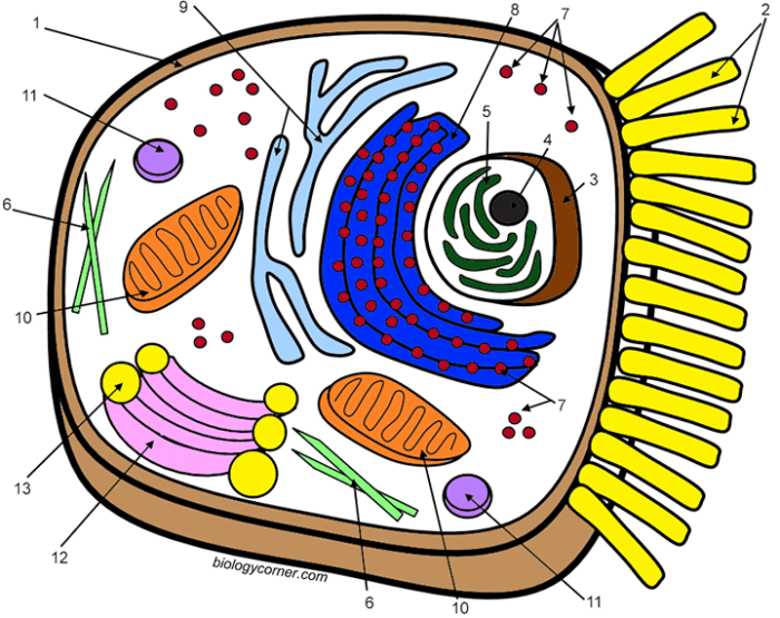
The coloring page should depict a simplified, yet accurate, representation of an animal cell. The cell membrane should be a distinct oval shape. Key organelles, such as the nucleus, mitochondria, ribosomes, endoplasmic reticulum (both rough and smooth), Golgi apparatus, lysosomes, and vacuoles, should be clearly drawn and labeled. Their relative sizes and positions within the cell should be approximately accurate.
For example, the nucleus should be relatively large and centrally located, while mitochondria should be smaller and scattered throughout the cytoplasm. The endoplasmic reticulum should be depicted as a network of interconnected membranes, differentiating between the rough ER (with ribosomes attached) and smooth ER.
Understanding animal cell structure is significantly enhanced through interactive learning activities like reading comprehension exercises and coloring. These activities become even more effective when paired with a detailed visual aid, such as a labeled diagram; for instance, you might find a helpful resource like this animal cell coloring key labeled diagram. Using such a key alongside your coloring and comprehension work allows for a more thorough and accurate understanding of the various organelles and their functions within the animal cell.
Choosing appropriate colors can further enhance understanding and memorability. Below is a suggested color palette:
- Cell Membrane: Light brown or beige. This represents the boundary of the cell, grounding the image.
- Nucleus: Dark purple. The nucleus is the control center, and purple signifies royalty and control.
- Nucleolus: Darker shade of purple. This visually differentiates the nucleolus within the nucleus.
- Mitochondria: Bright red or orange. Mitochondria are the “powerhouses” of the cell, and red/orange represents energy and activity.
- Ribosomes: Dark blue or teal. Ribosomes are small and numerous, and a darker, cooler color helps distinguish them from other organelles.
- Rough Endoplasmic Reticulum: Light blue. The ribosomes attached to the rough ER are already dark blue; the lighter blue background visually emphasizes the structure.
- Smooth Endoplasmic Reticulum: Light green. The smooth ER’s distinct function warrants a different color from the rough ER.
- Golgi Apparatus: Light yellow or gold. The Golgi apparatus is involved in packaging and transport, and yellow represents movement and transition.
- Lysosomes: Dark green. Lysosomes are involved in waste breakdown, and dark green can represent decomposition or recycling.
- Vacuoles: Light pink or peach. Vacuoles store materials, and the lighter, softer color represents containment.
- Cytoplasm: Light gray or pale yellow. This provides a background for the organelles, helping them stand out.
Animal Cell Crossword Puzzle
The crossword puzzle should include terms related to the organelles and their functions. For instance, clues could include: “Control center of the cell” (answer: NUCLEUS), “Powerhouse of the cell” (answer: MITOCHONDRIA), “Packages and transports proteins” (answer: GOLGI), “Site of protein synthesis” (answer: RIBOSOMES), “Digests waste materials” (answer: LYSOSOMES), “Cell’s outer boundary” (answer: MEMBRANE). The puzzle’s difficulty can be adjusted based on the students’ age and understanding.
The puzzle should be designed to be solvable, with a clear and logical layout.
Reinforcing Learning with Coloring and Crossword Activities
The coloring page and crossword puzzle can be used together to reinforce learning in several ways. Students can first complete the coloring page, focusing on the location and function of each organelle while associating them with the assigned colors. Then, they can attempt the crossword puzzle, applying their knowledge of the organelles and their functions. This dual approach strengthens memory through both visual and textual engagement.
The act of coloring helps to imprint the visual representation of the cell structure, while the crossword puzzle tests their understanding of the terminology and functions. This combination caters to diverse learning styles, leading to more effective and lasting retention of information.
Integrating Reading and Coloring: Animal Cell Reading Comprehension And Coloring

Combining reading comprehension exercises with coloring activities offers a multi-sensory approach to learning about animal cells, significantly enhancing knowledge retention and engagement. This integrated approach leverages the benefits of both textual and visual learning styles, catering to a wider range of learners.The simultaneous use of reading and coloring fosters a deeper understanding of animal cell structures and functions. Reading provides the factual information, while coloring acts as a visual reinforcement, helping to solidify the concepts learned.
This dual approach engages different parts of the brain, improving memory and comprehension.
Benefits of Visual Aids in Science Learning
Visual aids, such as coloring pages depicting animal cells with labeled organelles, are incredibly effective in simplifying complex scientific concepts. The act of coloring encourages active participation and focuses attention on specific details. For instance, coloring the nucleus a specific color and then reading about its function creates a strong association between the visual representation and the textual information, leading to better memorization.
Furthermore, visual aids are particularly helpful for visual learners, who process information more effectively through images and diagrams. They can also assist students with different learning styles and abilities.
Challenges and Solutions in Integrating Activities
Implementing integrated reading and coloring activities may present some challenges. One potential difficulty is ensuring that the coloring pages accurately reflect the information presented in the reading material. Discrepancies between the two could lead to confusion. To mitigate this, it’s crucial to carefully design the coloring pages to align precisely with the reading comprehension text. Another challenge could be managing the time allocated to both activities.
A solution is to plan the activities carefully, considering the age and learning abilities of the students, and providing clear instructions and time limits.
Adapting Activities for Different Learning Styles and Abilities
Adapting the activities to cater to diverse learning styles and abilities is essential for inclusive learning. For students who struggle with reading, the coloring activity can serve as a primary learning tool, supplemented by simplified reading materials or verbal explanations. Conversely, for students who prefer reading, the coloring activity can act as a supplementary tool to reinforce understanding.
Differentiation can also be achieved by providing different levels of complexity in the coloring pages, ranging from simple Artikels to detailed diagrams with numerous organelles to color and label. For students with fine motor skill challenges, larger coloring pages or alternative tactile activities, like building a 3D model of an animal cell, could be provided.
Helpful Answers
What are some alternative ways to use the coloring page besides coloring?
Students can label the organelles directly onto the coloring page, create a key identifying each organelle and its color, or use the page as a template for creating a 3D model of an animal cell.
How can I adapt these activities for older students?
For older students, increase the complexity of the reading passage and questions, incorporate more advanced concepts, and introduce research projects related to specific animal cells or cellular processes.
How can I assess student understanding after completing these activities?
Use the multiple-choice and short-answer questions provided, observe student participation and accuracy in labeling the coloring page, and conduct a follow-up quiz or class discussion.


