Organelle Descriptions for the Diagram

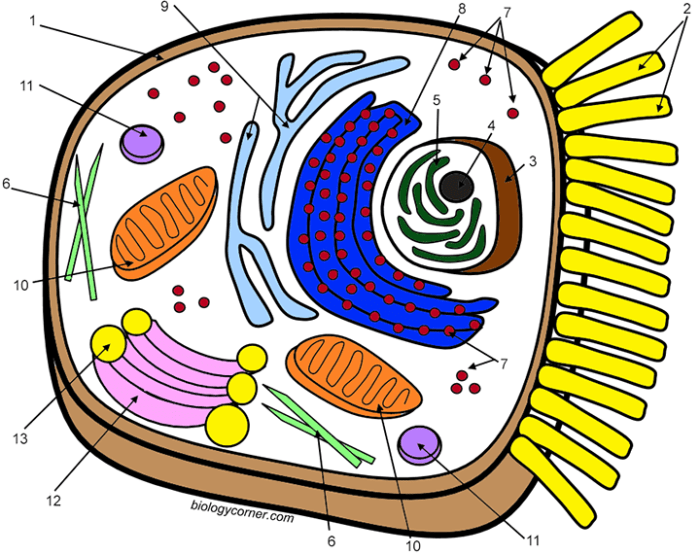
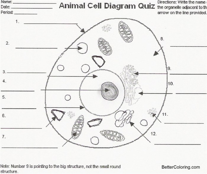
Animal cell coloring and labeling diagram – This section provides detailed descriptions of the key organelles found within a typical animal cell, focusing on their structure and function. Understanding these components is crucial to grasping the overall complexity and functionality of the cell. The descriptions below correlate directly with the organelles depicted in the accompanying diagram.
Nucleus, Animal cell coloring and labeling diagram
The nucleus is the control center of the cell, housing the cell’s genetic material, DNA. It’s a large, membrane-bound organelle typically located near the center of the cell. The nuclear membrane, or nuclear envelope, is a double membrane perforated by nuclear pores which regulate the transport of molecules in and out of the nucleus. Inside the nucleus, DNA is organized into chromosomes, and the nucleolus, a dense region within the nucleus, is responsible for ribosome biogenesis.
The nucleus dictates cellular activities by controlling gene expression, which determines which proteins are synthesized and when.
Mitochondria
Mitochondria are the powerhouses of the cell, responsible for generating most of the cell’s supply of adenosine triphosphate (ATP), the main energy currency. These double-membrane-bound organelles have a highly folded inner membrane called the cristae, which significantly increases the surface area for ATP production. The process of ATP generation is called cellular respiration, which involves a series of chemical reactions that break down glucose and other fuel molecules in the presence of oxygen.
The efficiency of mitochondria is critical for cell survival and function. Cells with high energy demands, like muscle cells, often contain many mitochondria.
Endoplasmic Reticulum
The endoplasmic reticulum (ER) is an extensive network of interconnected membranes extending throughout the cytoplasm. There are two main types: the rough endoplasmic reticulum (RER) and the smooth endoplasmic reticulum (SER). The RER is studded with ribosomes, giving it a rough appearance. It plays a key role in protein synthesis and modification. The SER lacks ribosomes and is involved in lipid synthesis, detoxification, and calcium storage.
The ER’s extensive network facilitates efficient transport of molecules within the cell.
Golgi Apparatus
The Golgi apparatus, also known as the Golgi complex or Golgi body, is a stack of flattened, membrane-bound sacs called cisternae. It acts as the cell’s processing and packaging center. Proteins and lipids synthesized by the ER are transported to the Golgi apparatus, where they are further modified, sorted, and packaged into vesicles for transport to other parts of the cell or for secretion outside the cell.
The Golgi apparatus plays a vital role in maintaining cellular organization and function.
Lysosomes
Lysosomes are membrane-bound organelles containing digestive enzymes. These enzymes break down waste materials, cellular debris, and foreign substances ingested by the cell through phagocytosis. Lysosomes maintain cellular cleanliness and prevent the accumulation of harmful substances. They are crucial for cellular recycling and defense against pathogens.
Ribosomes
Ribosomes are small, complex structures responsible for protein synthesis. They are composed of ribosomal RNA (rRNA) and proteins and can be found free in the cytoplasm or bound to the rough endoplasmic reticulum. Ribosomes translate the genetic code from messenger RNA (mRNA) into a sequence of amino acids, forming polypeptide chains that fold into functional proteins.
Cell Membrane
The cell membrane, also known as the plasma membrane, is a selectively permeable barrier that encloses the cell’s contents. It’s primarily composed of a phospholipid bilayer, with embedded proteins and cholesterol. The phospholipid bilayer provides a hydrophobic barrier, regulating the passage of substances into and out of the cell. Membrane proteins perform various functions, including transport, cell signaling, and cell adhesion.
Creating an accurate animal cell coloring and labeling diagram requires careful attention to detail. Understanding the functions of each organelle is key, and a helpful resource for verifying your work is readily available: you can check your answers against a comprehensive guide like the animal and plant cell coloring answer key. This will ensure your animal cell coloring and labeling diagram is both visually appealing and scientifically correct.
The cell membrane maintains the cell’s internal environment and facilitates interactions with its surroundings.
Cytoskeleton
The cytoskeleton is a complex network of protein filaments extending throughout the cytoplasm. It provides structural support, maintains cell shape, and facilitates intracellular transport. The cytoskeleton is composed of three main types of filaments: microtubules, microfilaments, and intermediate filaments. These filaments interact to provide dynamic structural support and enable cell movement and division.
Vacuoles
Animal cells contain small, temporary vacuoles involved in various functions such as storage, transport, and waste removal. Unlike plant cells, which typically have one large central vacuole, animal cell vacuoles are much smaller and less prominent. These vacuoles can store nutrients, water, or waste products temporarily before being transported or broken down.
Advanced Diagram Enhancements (Optional)

Taking your understanding of animal cells to the next level involves exploring their intricate structures and processes in greater detail. This section provides options for creating more advanced diagrams that showcase the complexity and functionality of the cell membrane, protein synthesis, and material transport. These enhancements will solidify your comprehension of cellular mechanisms.
Detailed Cell Membrane Diagram
A detailed depiction of the cell membrane should illustrate its fluid mosaic model. This model emphasizes the dynamic nature of the membrane, composed primarily of a phospholipid bilayer. The phospholipids, each with a hydrophilic head and two hydrophobic tails, arrange themselves to form a double layer, with the hydrophilic heads facing the aqueous environments inside and outside the cell, and the hydrophobic tails shielded within the membrane’s interior.
Interspersed within this bilayer are various proteins, including integral proteins that span the entire membrane and peripheral proteins that are loosely associated with either the inner or outer surface. Cholesterol molecules are also present, contributing to membrane fluidity and stability. Glycoproteins and glycolipids, carbohydrates attached to proteins and lipids respectively, extend from the outer surface and play roles in cell recognition and signaling.
A visually accurate representation would show these components in their relative proportions and spatial arrangements, conveying the dynamic and complex nature of the cell membrane.
Protein Synthesis Diagram
This diagram should illustrate the process of protein synthesis, starting from transcription in the nucleus to translation in the cytoplasm. The diagram should begin with DNA within the nucleus, highlighting the specific gene sequence being transcribed into messenger RNA (mRNA). The mRNA then exits the nucleus through nuclear pores and travels to a ribosome, either free in the cytoplasm or attached to the endoplasmic reticulum (ER).
The ribosome facilitates the translation of the mRNA sequence into a polypeptide chain, using transfer RNA (tRNA) molecules to bring the appropriate amino acids. The growing polypeptide chain folds into its three-dimensional structure, potentially undergoing further modifications within the ER and Golgi apparatus before reaching its final destination. Clear labeling of all involved molecules (DNA, mRNA, tRNA, ribosomes, amino acids, etc.) and cellular compartments is crucial for understanding the pathway.
Material Movement Across the Cell Membrane Diagram
This diagram should visually represent the different mechanisms of transport across the cell membrane. It should include depictions of passive transport (diffusion, osmosis, facilitated diffusion) and active transport (primary and secondary active transport, endocytosis, exocytosis). Passive transport should be shown as the movement of substances down their concentration gradients without energy expenditure. Active transport should be illustrated as the movement of substances against their concentration gradients, requiring energy in the form of ATP.
Specific examples of molecules transported via each mechanism should be included (e.g., oxygen diffusing into the cell, glucose transported via facilitated diffusion, sodium-potassium pump for active transport). The use of arrows to indicate the direction of movement and clear labels for each transport process is essential for clarity.
Plant and Animal Cell Comparison
| Organelle | Animal Cell Presence | Plant Cell Presence | Function |
|---|---|---|---|
| Cell Wall | Absent | Present | Provides structural support and protection |
| Chloroplasts | Absent | Present | Site of photosynthesis |
| Large Central Vacuole | Absent or small | Present | Maintains turgor pressure, stores water and nutrients |
| Cell Membrane | Present | Present | Regulates passage of materials into and out of the cell |
| Nucleus | Present | Present | Contains genetic material (DNA) |
| Mitochondria | Present | Present | Site of cellular respiration |
| Ribosomes | Present | Present | Protein synthesis |
| Golgi Apparatus | Present | Present | Modifies, sorts, and packages proteins |
| Endoplasmic Reticulum (ER) | Present | Present | Protein and lipid synthesis |
| Lysosomes | Present | Present (sometimes) | Waste breakdown and recycling |
Question Bank: Animal Cell Coloring And Labeling Diagram
What are the best tools for creating an animal cell diagram?
Many tools work well, from simple pencil and paper to digital drawing programs like Adobe Illustrator or even free online tools like Google Drawings. Choose a method comfortable for your skill level and desired level of detail.
How much detail should I include in my diagram?
The level of detail depends on your purpose. A basic diagram might show only major organelles, while a more advanced one could include subcellular structures. Aim for a balance between clarity and complexity.
Where can I find accurate images of animal cells for reference?
Reliable sources include scientific textbooks, reputable websites (e.g., those of universities or scientific organizations), and online databases of microscopy images.


